
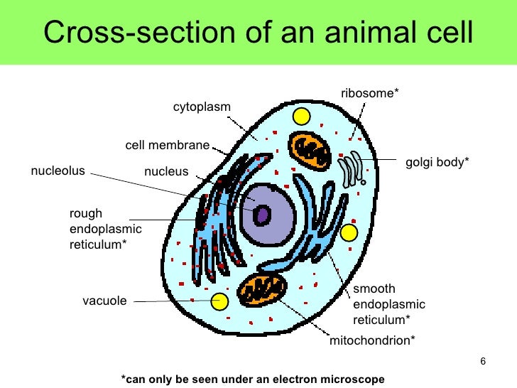
The cell membrane also known as plasma membrane or plasmalemma consists of three layers when viewed under the. The granulated area is the cell cytoplasm while the huge round part is the nucleus.
Transmission electron micrograph of animal cell Stock
Animal and plant cells have certain.
Animal cell under electron microscope. Most plant and animal cells are only visible under a light microscope, with dimensions between 1 and 100 micrometres. Animal cell diagram under electron microscope. Royalty free stock photos a typical cell labeled cell diagram animal cell cells worksheet.
Angelo on november 24, 2021. How to get a lion as a. In the given figure of an animal cell as observed under an electron microscope.
Animal cell diagram under electron microscope. Illustrate only a plant cell as seen under electron microscope. Human cheek cells are made of simple squamous epithelial cells, which are flat cells with a round visible nucleus that cover the inside lining of the cheek.c.
Cell 8 pictures of plant cells under a microscope plant cell structure under microscope plant and animal cells plant cell structure plant cell. The plant cell as more rigid and stiff walls. These are both specific types of cells, and from specific species.
The electron microscope two main advantages high resolving power (short wavelength of electrons) as electrons negatively are charged the beam can be focused using electromagnets as electrons are absorbed by molecules of air, a. A typical animal cell (as seen in an electron microscope) medical images for powerpoint. Below the basic structure is shown in the same animal cell, on the left viewed with the light microscope, and on the right with the transmission electron.
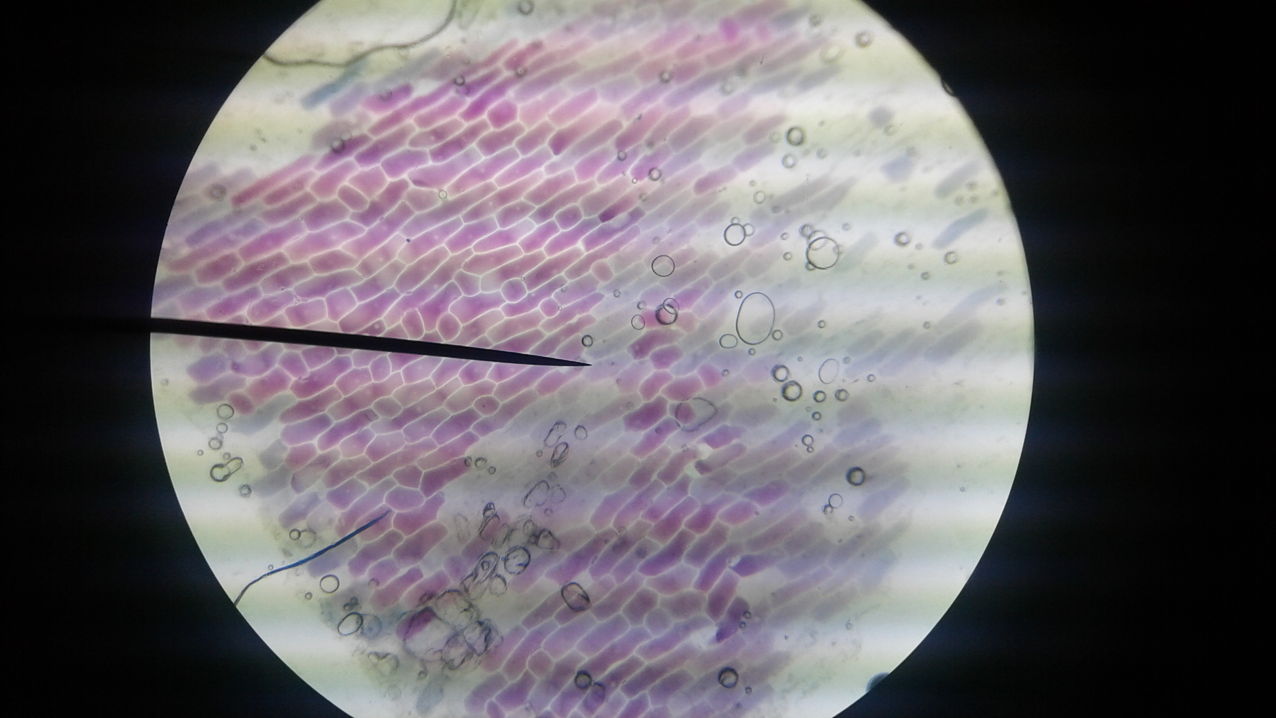
It was not until good light microscopes became available in the early part of the nineteenth century that all plant and animal tissues were discovered to be aggregates of individual cells. Within the cell, there is a shape of round with a circular structure of granulated part on the epithelial cells. You can observe this epithelial animal cell under microscope with high power.
Under a microscope, plant cells are surrounded by cell wall, which are not present in animal cells. Contents 1 when looking at plant and animal cells with an electron microscope you notice that the plant cells. The animal cell is more fluid or elastic or malleable in structure;
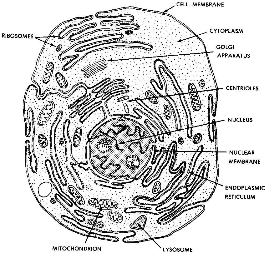
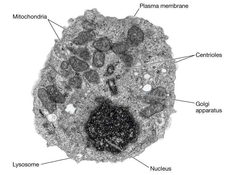
Typical animal cell pinocytotic vesicle lysosome golgi vesicles golgi vesicles rough er (endoplasmic reticulum) smooth er (no ribosomes) cell (plasma) membrane mitochondrion golgi apparatus nucleolus nucleus centrioles (2) each composed of 9. Here is an electron micrograph of an animal cell with the labels superimposed: We all remember that the human body is very intricate and one way i discovered to are aware of it is by means of the manner of human anatomy diagrams.
Eukaryotic is most complex cells consisting a true nucleus enclosed by a membrane. Admin send an email 3 weeks ago. A plant cell as seen under light microscope ultrastructure of animal and plant cells ultra structure of the cell is the fine structure of the cell as seen under electron microscope.
Below the basic structure is shown in the same animal cell, on the left viewed with the light microscope, and on the right with the transmission electron microscope. Angelo on august 20, 2021. Living cells cannot be observed using an.
The diagram is very clear and labeled. That’s the major difference between plant and animal cells under microscope. See more articles in category:
Illustrate only a plant cell as seen under an electron microscope. Compare an animal cell to a plant cell. Pin by nia on education plant cell electron microscope cell.
Cell organelles seen with light and electron microscopes animal cell under light microscope plant cell under light microscope. Diagram of animal cell under electron microscope. The fine structure of a cell is revealed by electron microscopy for the highest magnification and best resolution, one must turn to an electron microscope, which can reveal details down to a few nanometers.
There are two categories of cells, eukaryotic and prokaryotic. Year 11 bio key points cell organelles and their function animal cell cell organelles eukaryotic cell. Plant cell diagram electron microscope.
You see that many features are in common. Generalized cell is used for structure of animal cell and plant cell to present the common parts, appearing in various parts of the bodies of animals and plants. We have got 7 pic about diagram of animal cell seen under electron microscope images, photos, pictures, backgrounds, and more.
Structure of plant and animal cells under an electron microscope. Animal cells have a basic structure. Plant, animal and bacterial cells have smaller components each with a specific function.
A cell is the smallest functional and structural entity of life that it is easier observing animal cell under light microscope. Generalized cell is used for structure of animal cell and plant cell to present the. They are all typical elements of a cell.
How is it different from an animal cell? There are one or more cells that form organism. The electron microscope electron microscopes use a beam of electrons instead of beams or rays of light.
Cell Structure and Organisation

Cell Theory Biology 102 Basic Units of Life
Images 01. Introduction and Terminology Basic Human Anatomy

labeled animal cell under electron microscope 8745961 orig
animal cell microscope Biological Science Picture

Eukaryotic Animal Cell Under Microscope Micropedia

Scanning Microscopy, Confocal Micrography and FlatBed

Q14 Draw a large diagram of an animal cell as seen through

bacterial growth petri dish Tìm với Google Ocean
Edexcel IAL Biology 2.3.3 Describe the ultrastructure of
Pictures Of Animal Cells Under A Microscope Micropedia

Q14 Draw a large diagram of an animal cell as seen through

Animal Cells and Plant Cells Cell As a Unit of Life
Electron Microscope Eukaryotic Animal Cell Micropedia


Rana ray diagram of animal cell seen through electron

labeled animal cell under electron microscope midbodyl

Draw a large daigram of an animal cell as seen through an

cellfig10.jpg (1378×1080) Scanning electron microscope

Transmission Electron Microscopy PinBio_1002.2016