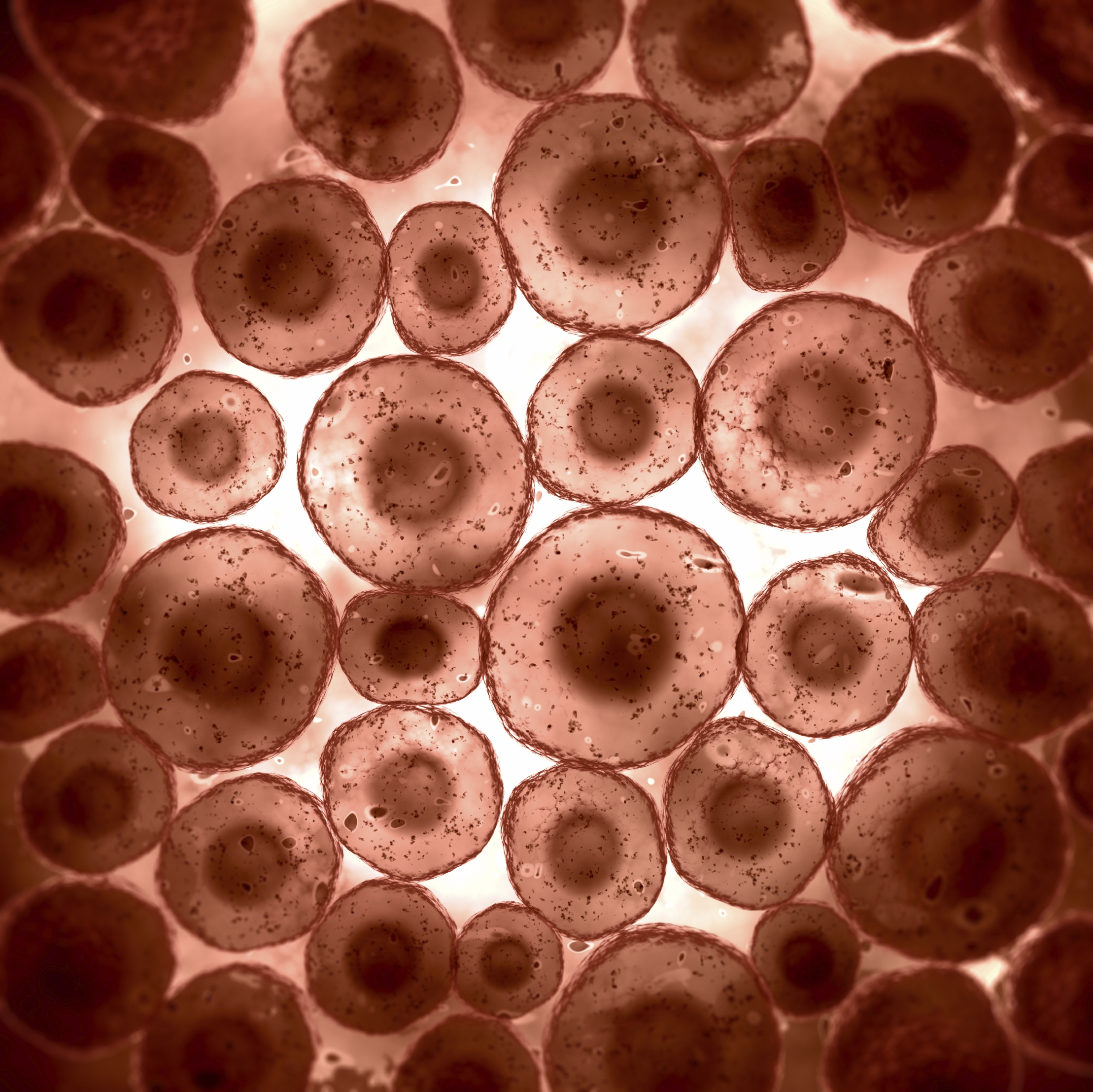
Microscopically, animal cells from the same tissue of an animal will have varied sizes and shapes due to the lack of a rigid cell wall. Angelo on august 20, 2021.
Pictures Of Animal Cells Under A Microscope Micropedia
Viewing animal cells under a microscope.

Animal cell under microscope. Animal cell diagram under electron microscope. Browse 4,547 animal cells under microscope stock photos and images available or start a new search to explore more stock photos and images. Structures viewed under an optical microscope can be measured using the formula:
Beneath a plant cell's cell wall is a cell membrane. Under the microscope, animal cells appear different based on the type of the cell. The diagram is very clear and labeled.
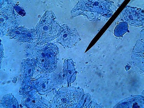
Within the cell, there is a shape of round with a circular structure of granulated part on the epithelial cells. Cell nuclei of amphibian larva seen under a microscope, at x750 magnification. Year 11 bio key points cell organelles and their function animal cell cell organelles eukaryotic cell.
You can observe this epithelial animal cell under microscope with high power. Royalty free stock photos a typical cell labeled cell diagram animal cell cells worksheet. There are one or more cells that form organism.
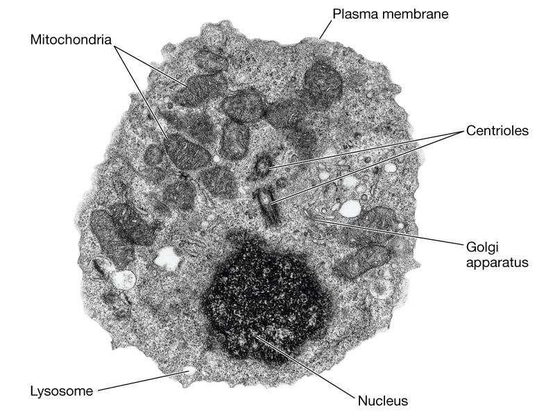
There are two categories of cells, eukaryotic and prokaryotic. In the given figure of an animal cell as observed under an electron microscope. But at the same time it is interpretive.
Under a microscope, plant cells from the same source will have a uniform size and shape. Eukaryotic is most complex cells consisting a true nucleus enclosed by a membrane. Magnification = size of image / size of real object structure of animal cell and plant cell under microscope + diagrams learn the structure of animal cell and plant cell under light microscope.
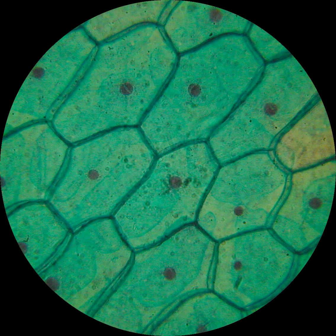
Viewing animal cells under a microscope. Human cheek cells are made of simple squamous epithelial cells, which are flat cells with a round visible nucleus that cover the inside lining of the cheek.c. In this way, what does a animal cell look like under a microscope?
Epithelial cells surround the internal surface of the mouth which can be taken out using finger nails or a small spoon. Nucleus, cytoplasm, cell membrane, chloroplasts and cell wall (last 2 organelles are only present in plant cells). Here's a diagram of a plant cell:
In the given figure of an animal cell as observed under an electron microscope. Bookfanatic89 diagram of plant cell under electron microscope. Compare an animal cell to a plant cell.
Animal cells almost all animals and plants are made up of cells. What parts of an animal cell can you see under a light microscope? Microscopically, animal cells from the same tissue of an animal will have varied sizes and shapes due to the lack of a rigid cell wall.
Unlike the eukaryotic cells of plants and fungi, animal cells do not have a cell wall. Cell 8 pictures of plant cells under a microscope plant cell structure under microscope plant and animal cells plant cell structure plant cell. What type of microscope is needed to see a plant cell?
Meiosis cell division 3d cell cellular division embryo 3d cell animal embryo reproductive health blood cells under microscope the cell cytyoplasm cytoplasm. An animal cell represents an eukaryotic cell in which true nucleus and other membrane bound organelles such as mitochondria. See animal cell under microscope stock video clips.
Diagram of animal cell under electron microscope. It was not until good light microscopes became available in the early part of the nineteenth century that all plant and animal tissues were discovered to be aggregates of individual cells. For organelles that can be seen under the light microscope are mainly the protoplasm:
That’s the major difference between plant and animal cells under microscope. Contents 1 when looking at plant and animal cells with an electron microscope you notice that the plant cells. You know, animal cell structure contains only 11 parts out of the 13 parts you saw in the plant cell diagram, because chloroplast and cell wall are available only in a plant cell.
Browse 231 animal cells under microscope stock photos and images available, or start a new search to explore more stock photos and images. Most of the cells are microscopic in size and can only be seen under the microscope. So it is important to note that.
The diagram is very clear, and labeled; Under a microscope, plant cells from the same source will have a uniform size and shape.beneath a plant cell's cell wall is a cell membrane. There are also more intriguing shapes such as curved, spherical, concave and rectangular.
Most cells, both animal and plant, range in size between 1 and 100 micrometers and are thus visible only with the aid of a microscope. A cell is the smallest functional and structural entity of life that it is easier observing animal cell under light microscope. Animal cell diagram under electron microscope.
Below the basic structure is shown in the same animal cell, on the left viewed with the. Angelo on november 24, 2021. Diagram of animal cell under electron microscope.
Pin by nia on education plant cell electron microscope cell.

Smart Science Pro December 2011
Pictures Of Animal Cells Under A Microscope Micropedia
Plant & Animal Cells Staining Lab Answers Online
Animal cell microscope picture Animal cells under
Animal Cells Under Microscope Stock Photo Download Image


microscopic images of plant and animal cells Google

A school of fish Plant and Animal Cells through the
Animal cells under microscope. — Stock Photo © vladnikon

animal cells under a microscope Under the microscope

Plant and Animal Cells
Edexcel IAL Biology 2.3.3 Describe the ultrastructure of
Animal Cells Under Microscope Stock Photo Download Image

animal cell under a microscope
Animal Cells and Plant Cells Cell As a Unit of Life
animal cell microscope Biological Science Picture

Cells under a microscope Biological Science Picture
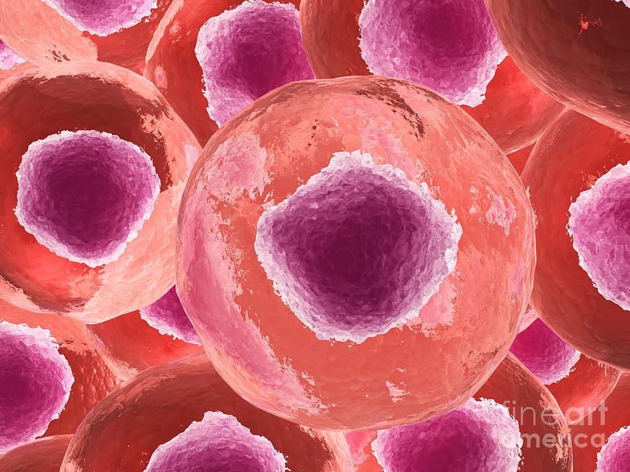
Microscopic View Of Animal Cell Digital Art by Stocktrek
Microscope Human Cell Images Micropedia

Animal Cells Under Light Microscope Micropedia