
Plant, animal and bacterial cells have smaller components each with a specific function. We all remember that the human body is very intricate and one way i discovered to are aware of it is by means of the manner of human anatomy diagrams.
A Eukaryotic Cell in its basic form. prokaryotic vs
Coloured scanning electron micrograph (sem) of sensory hair cells from the inner ear.
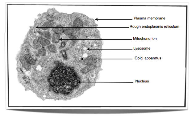
Electron micrograph of animal cell. Animal and plant cells have certain. Plant, animal and bacterial cells have smaller components each with a specific function. But at the same time it is interpretive.
These are animal cells that are specialized to secrete large quantities of digestive enzymes. The electron micrograph of nucleus: If you have a really good microscope and an excellent preparation you may be able to.
(1) nucleus was discovered by brown (1831). Plant cell diagram electron microscope the greatest garden plant cell diagram animal cell structure cell diagram. Cell structure light and electron microscopes allow us to see inside cells.
Animal cell electron micrograph labeling. Transmission electron micrograph (tem) of lysosomes. Learn vocabulary, terms, and more with flashcards, games, and other study tools.
Plant cell diagram electron microscope. The electron micrograph displayed below illustrates many of the plant cell characteristics discussed the cell wall, large central vacuole and chloroplasts are clearly visible also visible is the clearly defined nucleus containing chromatin nucleus chromatin the vacuole in this mature plant cell from a leaf is large, and occupies about 80% of This is an electron micrograph of nucleus.
Taking up most of the cell is the nucleus, where genes are stored in the form of chromosomes. Cell structure human cell diagram cell diagram animal cell drawing. The movement is converted to an electrical signal that is passed on to the brain.
The following sizes are ranges only. Electron micrograph animal cell under electron microscope. The diagram is very clear, and labeled;
Transmission electron micrograph of a mammalian tissue culture cell. They will have all the organelles of an animal cell but will have many ribosomes and rough er to create the enzymes which are proteins and transport them outside the cell. Cell 8 pictures of plant cells under a.
(2) it is a characteristic entity of almost all eukaryotic cells except mammalian rbcs. Start studying aice biology chapter 1: Holes in the membrane (at right, for example) allow large molecules to pass out into the cell cytoplasm.
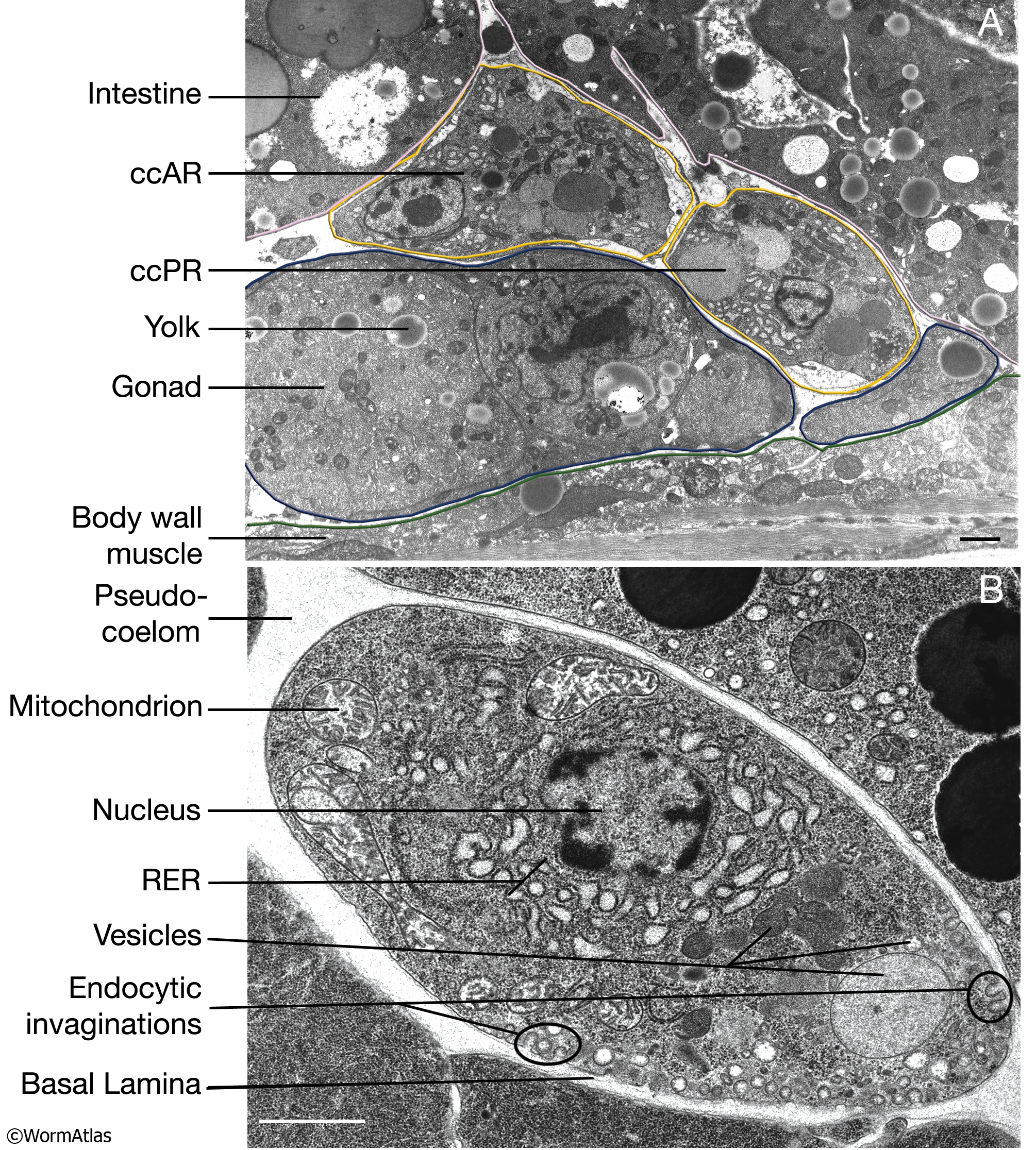
Figure 21 epithelial cells often display extensive basal plasma membrane infoldings as observed in this electron micrograph: I pinimg com 474x d7 30 97 d730978ce46b9f67e308. The dark zone (centre right) in the nucleus is the nucleolus.
This is the most active part of the nucleus, and contains unravelled chromosomes involved in making. Angelo on august 20, 2021. They are spherical vesicles which contain hydrolytic enzymes that can break down virtually all kinds of biomolecules (waste.
An electron micrograph of a mouse liver cell dna learning center electrons cell learning centers. You may also find mitochondrion, cell wall, an algal cell, animal cell, plasma membrane, cytoplasm, nucleolus, rough endoplasmic reticulum, lysosome in this image. Heterochromatin figure 22 a cell in mitosis (metaphase):
The dark grey circles above the nucleus are mitochondria, where fats and sugars are. Plant cells tem electron micrograph of a plant cell showing key features You should be able to describe and interpret photomicrographs, electron micrographs and drawings of typical animal cells.
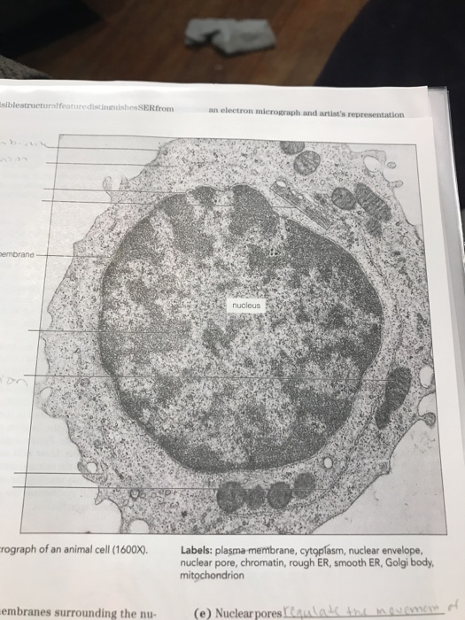
Calculate the magnification used to view the image. These cells are surrounded by a fluid called endolymph. The actual diameter of the animal cell is 45 um.) the observed diameter of the animal cell in the electron micrograph is 1.8 cm.
Transmission electron micrograph of part of a mouse liver cell. The diagram is very clear and labeled. Plant cells are often larger than animal cells.
Pin by nia on education plant cell electron microscope cell. Diagram of animal cell under electron microscope. Chromosomes figure 23 a cell in mitosis (telophase):
As sound enters the ear it causes waves to form in the endolymph, which in turn cause the hairs to move. Actual cell size may be larger or smaller and will depend largely on cell type. Here's a diagram of a plant cell:
They have many mitochondria to supply the atp needed for these processes. This is a colored scanning electron micrograph of human red and white blood cells. Transmission electron micrograph (tem) of lysosomes.
Animal cell diagram under electron microscope. Seen under the electron microscope (not to scale) one function of organelle cell type(s) in which organelle is located mitochondrion animal and plant assemble microtubules to produce the mitotic spindle rough endoplasmic reticulum protein synthesis golgi apparatus animal and plant photosynthesis plant only [8] [total: They are spherical vesicles which contain hydrolytic enzymes that can break down virtually all kinds of biomolecules (waste.
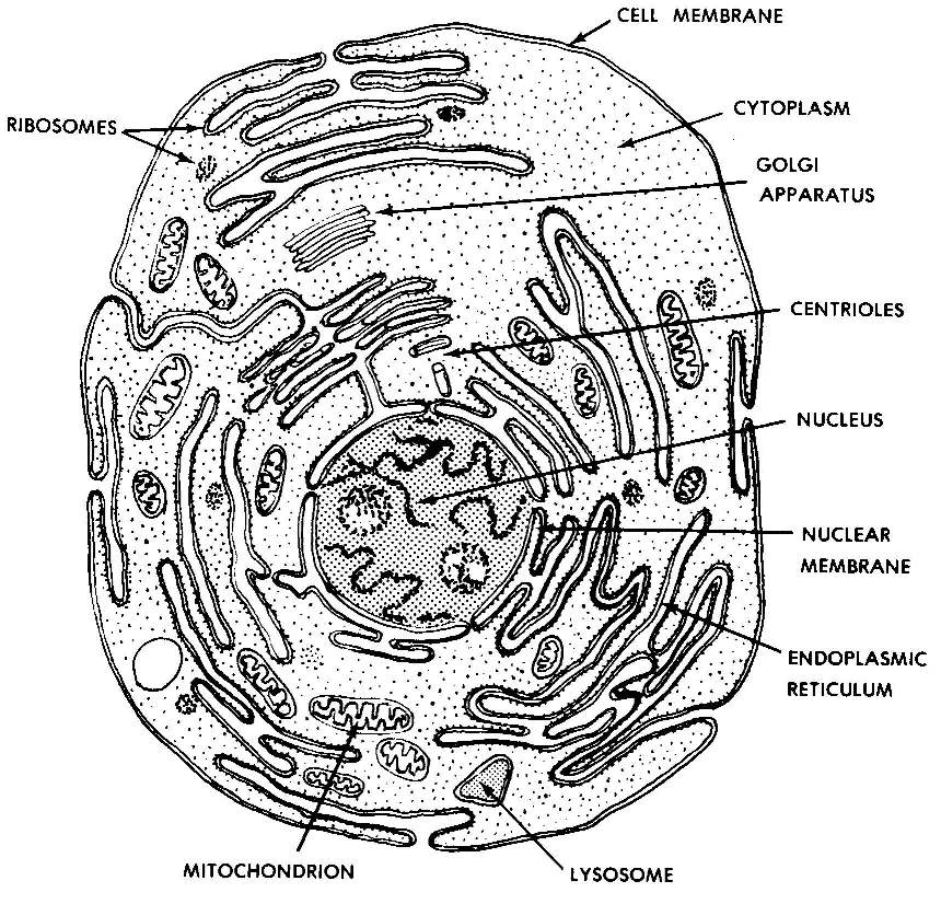
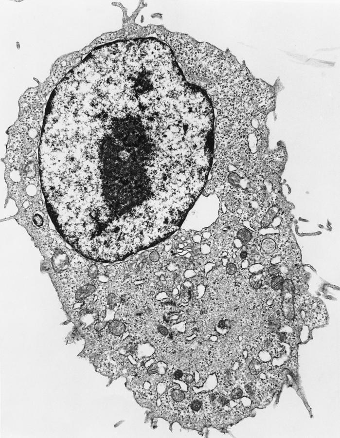
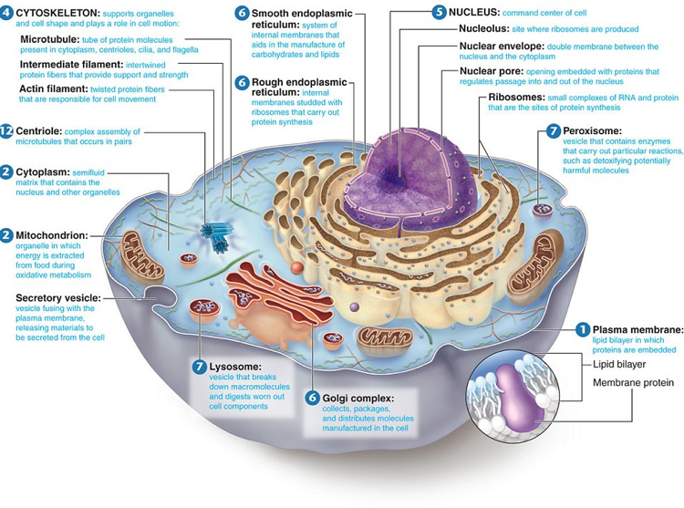
In the lower frame is the cell nucleus, bound by a delicate nuclear membrane. A typical animal cell (as seen in an electron microscope) medical images for powerpoint 1. Typical animal cell pinocytotic vesicle lysosome golgi vesicles golgi vesicles rough er (endoplasmic reticulum) smooth er (no ribosomes) cell (plasma) membrane mitochondrion golgi apparatus nucleolus nucleus centrioles (2) each composed of 9.
In this image, you will find plant cell and the animal cell under electron micrograph, peroxisome, nucleus, vacuole, chloroplast, golgi complex in it. Determine the sizes of the animal cell, the plant cell and the chloroplast in the electron micrographs provided. Year 11 bio key points cell organelles and their function animal cell cell organelles eukaryotic cell.
Images 01. Introduction and Terminology Basic Human Anatomy
Tem Of Animal Cell Photograph by Dr Gopal Murti
Electron Microscope Eukaryotic Animal Cell Micropedia

Transmission Electron Micrograph Of An Animal Cell

Plant Cell And Animal Cell Under Electron Micrograph

Cell Theory Biology 102 Basic Units of Life
Year 11 Bio. Key Points Cell organelles and their function
.jpg)
CcFIG 5 Legend
Cellular portraits — Opuntia Visual

Solved Label The Ectron Micrograph Of An Animal Cell. Inc

Draw a large daigram of an animal cell as seen through an

Electron Microscope Eukaryotic Animal Cell Micropedia
![]()
Transmission Electron Micrograph Of Animal Cell Photograph

Rosa Rubicondior Ungodly Complexity
IB Biology Notes 2.3 Eukaryotic cells

labeled animal cell under electron microscope midbodyl

Animal Cells and Plant Cells Cell As a Unit of Life
Oncology Basics 2016 Understanding the Cells
animal cell microscope Biological Science Picture
